Archive for February, 2019
February 27, 2019
A larva/nymph in the Family Gomphidae (Clubtails) was collected by Bob Perkins from the New River in southwestern Virginia. The larva died before it metamorphosed into an adult.

“Generic Gomphid” larva (preserved specimen) | New River, VA USA
An Ashy Clubtail dragonfly nymph was also collected by Bob Perkins. (The date and location where the specimen was collected are unknown.) The nymph was reared in captivity until it emerged on 21 March 2017 and metamorphosed into an adult female. This specimen is the exuvia from the nymph.
Although face-head of the “Generic Gomphid” and Ashy Clubtail look similar, they aren’t identical. More later after the specimen is keyed out carefully.
Related Resources
Tech Tips
13 photos were used to create the focus stack of the “Generic Gomphid.” A single focus point was positioned over select anatomical features; photos were taken at each point of interest.
The following equipment was used to shoot the composite image of the “Generic Gomphid”: Canon EOS 5D Mark II digital camera, in manual mode; Canon MP-E 65mm Macro lens (set for f/11 at ~2.5x); a Canon MT-26EX-RT Macro Twin Lite set for “Master” mode, and a single external flash set for “Slave” mode — a Godox TT685C Thinklite TTL Flash fitted with a Lastolite Ezybox Speed-Lite 2 flash modifier.
Auto power-off was disabled for the camera and external flash units.
Adobe Photoshop CC 2017 was used to create the focus stack, as well as spot-heal and sharpen the final output.
Copyright © 2019 Walter Sanford. All rights reserved.
Tags:Ashy Clubtail dragonfly, Family Gomphidae (Clubtails), gear talk, high-speed sync, Phanogomphus lividus, studio photography
Posted in Aperture, Canon EOS 5D Mark II, Canon MP-E 65mm Macro lens, Canon MT-26EX-RT Macro Twin Lite, digital photography, dragonflies, Godox TT685C, How To, Lastolite flash modifier, macro photography, natural science, Photoshop, wildlife photography | 2 Comments »
February 25, 2019
This post features a focus-stacked composite image that shows a dorsal view of an odonate larva/nymph from the Family Cordulegastridae (Spiketails) that was collected and reared by Bob Perkins. The larva died before it metamorphosed into an adult.

Cordulegaster sp. larva (female) | dorsal view
Most larvae go through 10-13 stages of development known as “instars.” The author lacks sufficient experience to identify the instar of this specimen, although it appears to be one of the later stages as indicated by its well-developed wing pads.
Related Resources
Tech Tips
12 photos were used to create the focus stack. A single focus point was positioned over select anatomical features; photos were taken at each point of interest.
The following equipment was used to shoot all of the photographs for the focus-stacked composite image, shown above: Canon EOS 5D Mark II digital camera, in manual mode; Kenko 20mm macro automatic extension tube; Canon EF100mm f/2.8L Macro lens (set for manual focus); and Canon MT-26EX-RT Macro Twin Lite set for “Master” mode, and several external flashes set for “Slave” mode including Canon 580 EX- and Canon 580EX II Speedlites and a Godox TT685C Thinklite TTL Flash fitted with a Lastolite Ezybox Speed-Lite 2 flash modifier.
Auto power-off was disabled for the camera and all external flash units.
Adobe Photoshop CC 2017 was used to create the focus-stacked composite image, as well as spot-heal and sharpen the final output.
Copyright © 2019 Walter Sanford. All rights reserved.
Tags:Family Cordulegastridae (Spiketails), gear talk, high-speed sync, larva, nymph, studio photography
Posted in Aperture, Canon 580EX II Speedlite, Canon 580EX Speedlite, Canon EF 100mm Macro lens, Canon EOS 5D Mark II, Canon MT-26EX-RT Macro Twin Lite, digital photography, dragonflies, extension tubes, Godox TT685C, How To, Lastolite flash modifier, macro photography, natural science, Photoshop, wildlife photography | 2 Comments »
February 22, 2019
Bob Perkins collected and reared an odonate larva/nymph from the Family Cordulegastridae (Spiketails). The larva died before it metamorphosed into an adult.
This post features a focus-stacked composite image that shows a ventral view of the preserved larva; a composite image showing the dorsal view will be published in my next blog post.

Cordulegaster sp. larva (female) | ventral view
This individual is a female, as indicated by her rudimentary ovipositor that can be seen on the ventral side of the specimen along the boundary between abdominal segments eight and nine (S8-9). Do you see it?
Related Resources
Tech Tips
Nine (9) photos were used to create the focus stack. A single focus point was positioned over select anatomical features; photos were taken at each point of interest.
The following equipment was used to shoot all of the photographs for the focus-stacked composite image, shown above: Canon EOS 5D Mark II digital camera, in manual mode; Kenko 20mm macro automatic extension tube; Canon EF100mm f/2.8L Macro lens (set for manual focus); and Canon MT-26EX-RT Macro Twin Lite set for “Master” mode, and several external flashes set for “Slave” mode including Canon 580 EX- and Canon 580EX II Speedlites and a Godox TT685C Thinklite TTL Flash fitted with a Lastolite Ezybox Speed-Lite 2 flash modifier.
Auto power-off was disabled for the camera and all external flash units.
Adobe Photoshop CC 2017 was used to create the focus-stacked composite image, as well as spot-heal and sharpen the final output.
Copyright © 2019 Walter Sanford. All rights reserved.
Tags:Family Cordulegastridae (Spiketails), gear talk, high-speed sync, larva, nymph, ovipositor, studio photography
Posted in Aperture, Canon 580EX II Speedlite, Canon 580EX Speedlite, Canon EF 100mm Macro lens, Canon EOS 5D Mark II, Canon MT-26EX-RT Macro Twin Lite, digital photography, extension tubes, Godox TT685C, How To, Lastolite flash modifier, macro photography, natural science, Photoshop, wildlife photography | 1 Comment »
February 20, 2019
…and smell, er, see the roses. Seriously, man doesn’t live by odonates alone!
The following gallery features several photos of a Valentines Day gift of appreciation from The Beacon of Groveton. Nearly a week after the red rose was delivered, it’s beginning to show a little “character.”

19 FEB 2019 | BoG Photo Studio | Valentines Day rose

19 FEB 2019 | BoG Photo Studio | Valentines Day rose

19 FEB 2019 | BoG Photo Studio | Valentines Day rose
Tech Tips
The subject was posed in front of a black background — the lower door of the refrigerator!
The following equipment (shown below) was used to shoot the preceding photos: Fujifilm X-T1 digital camera; Fujifilm MCEX-16 extension tube; Fujinon XF80mm macro lens; Godox XProF TTL Wireless Flash Trigger for Fujifilm cameras; and a Godox TT685C Thinklite TTL Flash for Canon Cameras fitted with a Lastolite Ezybox Speed-Lite 2 flash modifier. A Sunpak LED-160 Video Light was used to add fill light to some of the photos.
Adobe Photoshop CC 2017 was used to spot-heal and sharpen the final output.
Copyright © 2019 Walter Sanford. All rights reserved.
Tags:gear talk, studio photography
Posted in Aperture, digital photography, extension tubes, Fujifilm X-T1, Fujinon XF80mm macro lens, Godox TT685C, Godox XProF, How To, Lastolite flash modifier, macro photography, natural science, Photoshop, Sunpak LED-160, wildlife photography | 3 Comments »
February 18, 2019
A larva/nymph in the Family Corduliidae (Emeralds) was collected by Bob Perkins on 02 December 2017 from a pond in Orange Park, Florida (USA). The larva died before it metamorphosed into an adult.
As you can see by looking at a close-up image of the face-head at 3x magnification, there is no horn on the face of the specimen. Therefore this individual is not a member of Family Macromiidae (Cruisers), as I speculated in my last blog post.

“Generic Baskettail” larva (preserved specimen) | face-head
Knowing the limits of our expertise
Although I still need to key out the specimen carefully, at this point I’m certain Bob is correct — the larva is a member of the Family Corduliidae (Emeralds). The question that remains unanswered is “Which genus/species?” We may never know the answer, as Bob and I have reached the limit of our experience and expertise.
I did a quick scan of Paulson’s [book], looking at the Emerald Family. Here, according to the range maps, are the possibilities for Orange Park [FL]. I believe you can see why I stopped at “generic basketttail.” Source Credit: Bob Perkins.
What do you think the identity is? Most of the items in the preceding species list feature links to photos of odonate larvae/exuviae. See the links to BugGuide from the scientific names in the list.
Related Resource: Test shots: “Generic Baskettail?”
Tech Tips
Four (4) photos were used to create the preceding focus-stacked composite image. A single focus point was positioned over the face, between the antennae. At a magnification ratio of 3:1, it’s difficult to manually focus on a single point — the slightest movement around the macro rig changes focus unintentionally. A simple work-around for this problem is to take several shots of the same focus point and create a composite image of the photos.
The following equipment was used to shoot the preceding composite image: Canon EOS 5D Mark II digital camera, in manual mode; Canon MP-E 65mm Macro lens (set for f/16 at 3x); a Canon MT-26EX-RT Macro Twin Lite set for “Master” mode, and a single external flash set for “Slave” mode — a Godox TT685C Thinklite TTL Flash fitted with a Lastolite Ezybox Speed-Lite 2 flash modifier. A Sunpak LED-160 Video Light was used to add fill light to the top of the subject.
Auto power-off was disabled for the camera and external flash units.
Adobe Photoshop CC 2017 was used to create the focus stack, as well as spot-heal and sharpen the final output.
Copyright © 2019 Walter Sanford. All rights reserved.
Tags:Family Corduliidae (Emeralds), gear talk, larva, nymph
Posted in Aperture, Canon EOS 5D Mark II, Canon MP-E 65mm Macro lens, Canon MT-26EX-RT Macro Twin Lite, digital photography, dragonflies, Godox TT685C, How To, Lastolite flash modifier, macro photography, natural science, Photoshop, Sunpak LED-160, wildlife photography | 1 Comment »
February 15, 2019
A larva/nymph in the Family Corduliidae (Emeralds) was collected by Bob Perkins on 02 December 2017 from a pond in Orange Park, Florida (USA). The larva died before it metamorphosed into an adult.
Test shots of this beautifully preserved specimen were taken using a small-ish aperture of f/11 for greater depth of field. The following photos are “one-offs,” that is, not composite images.
Dorsal
A single focus point — located on the thorax (specifcally, the “shoulder pad” along the right side of the body) — was used to shoot this photo. The specimen has enough “relief” that focus on the wing pads and dorsal hooks is slightly soft. This view of the larva is a good candidate for focus-stacking.
The terminal appendages (cerci, epiproct, paraprocts) are shown clearly in the following photo.

“Generic Baskettail” larva (preserved specimen) | Orange Park, FL USA
Bob’s best guess of the identity of the specimen is Epitheca sp., either Common Baskettail (Epitheca cynosura) or Prince Baskettail (Epitheca princeps).
Whenever I see an odonate larvae/exuviae with long legs, my first thought is Family Macromiidae (Cruisers). Then I check for a horn on top of the head, a key field mark for Cruisers. Look closely at the dorsal view of the larva and I think you’ll agree with me there appears to be a horn on the head. I would like to take close-up photos of the head and key out the specimen in order to determine its identity. In the meantime, my best guess is Stream Cruiser (Didmops transversa) as indicated by the lateral spines on abdominal segment nine (S9) and the absence of a dorsal hook on S10.
Ventral
The ventral side of the specimen has almost no “relief,” so a “one-off” focused on the thorax looks fairly good from head-to-tail.

“Generic Baskettail” larva (preserved specimen) | Orange Park, FL USA
Related Resource: “Generic Baskettail” (definitely not a Cruiser)
Tech Tips
The following equipment (shown below) was used to shoot the preceding photos: Fujifilm X-T1 digital camera; Fujifilm MCEX-16 extension tube; Fujinon XF80mm macro lens; Godox XProF TTL Wireless Flash Trigger for Fujifilm cameras; Godox TT685F Thinklite TTL Flash for Fujifilm Cameras; Godox TT685C Thinklite TTL Flash for Canon Cameras fitted with a Lastolite Ezybox Speed-Lite 2 flash modifier; and a Canon 580EX II Speedlite mounted on a Godox X1R-C TTL Wireless Flash Trigger Receiver for Canon. A new Godox TT685O Thinklite TTL Flash for Olympus/Panasonic Cameras was added to an array of radio-controlled external flash units used to light the specimen. All flashes were set for Manual Mode at 1/128 power.
Adobe Photoshop CC 2017 was used to spot-heal and sharpen the final output.
Copyright © 2019 Walter Sanford. All rights reserved.
Tags:cerci, Common Baskettail dragonfly, dorsal hooks, epiproct, Epitheca cynosura, Epitheca princeps, Family Corduliidae (Emeralds), larva, nymph, paraprocts, Prince Baskettail dragonfly, terminal appendages
Posted in Aperture, Canon 580EX II Speedlite, digital photography, dragonflies, extension tubes, Fujifilm X-T1, Fujinon XF80mm macro lens, Godox TT685C, Godox TT685F, Godox TT685o/p, Godox X1R-C, Godox XProF, How To, Lastolite flash modifier, macro photography, natural science, Photoshop, wildlife photography | 1 Comment »
February 13, 2019
“It’s like Deja vu all over again.” (Source Credit: Yogi Berra.) But seriously folks, if you were thinking “Hey these pictures look familiar!” then you’re right. My last blog post features “one-offs” of the same subject, that is, photos with a single focus point on the mid-body of the specimen.
This post features focus-stacked composite images that show dorsal- and ventral views of a preserved larva in the Family Gomphidae (Clubtails) that was collected by Bob Perkins. I think both composite images look better than the “one-offs” in my last blog post; the difference is especially noticeable by looking at the head and tail in the vernal view.
Dorsal
Five (5) photos were used to create the first focus stack. A single focus point was positioned over select anatomical features; photos were taken at each point of interest.

“Generic Gomphid” larva (preserved specimen) | New River, VA USA
Ventral
Six (6) photos were used to create the second focus stack.

“Generic Gomphid” larva (preserved specimen) | New River, VA USA
Related Resources
Tech Tips
The following equipment (shown below) was used to shoot the preceding photos: Fujifilm X-T1 digital camera; Fujifilm MCEX-16 extension tube; Fujinon XF80mm macro lens; Godox XProF TTL Wireless Flash Trigger for Fujifilm cameras; Godox TT685F Thinklite TTL Flash for Fujifilm Cameras; Godox TT685C Thinklite TTL Flash for Canon Cameras fitted with a Lastolite Ezybox Speed-Lite 2 flash modifier; and a Canon 580EX II Speedlite mounted on a Godox X1R-C TTL Wireless Flash Trigger Receiver for Canon. A new Godox TT685O Thinklite TTL Flash for Olympus/Panasonic Cameras was added to an array of radio-controlled external flash units used to light the specimen. All flashes were set for Manual Mode at 1/128 power.
Adobe Photoshop CC 2017 was used to create the preceding focus-stacked composite images, as well as spot-heal and sharpen the final output.
Copyright © 2019 Walter Sanford. All rights reserved.
Tags:Family Gomphidae (Clubtails), gear talk, studio photography
Posted in Aperture, Canon 580EX II Speedlite, digital photography, dragonflies, extension tubes, Fujifilm X-T1, Fujinon XF80mm macro lens, Godox TT685C, Godox TT685F, Godox TT685o/p, Godox X1R-C, Godox XProF, How To, Lastolite flash modifier, macro photography, natural science, Photoshop, wildlife photography | 1 Comment »
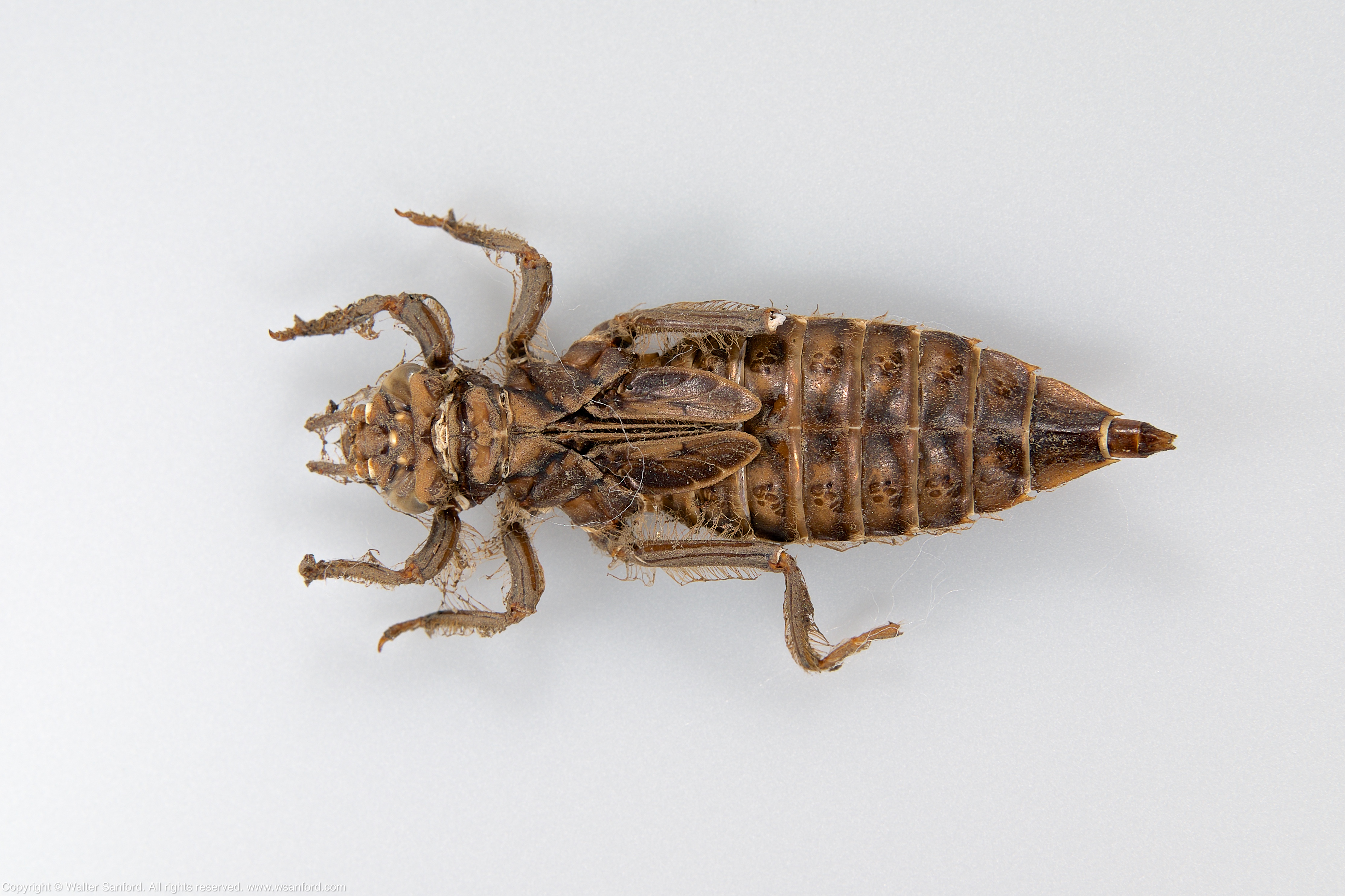
February 11, 2019
A larva/nymph in the Family Gomphidae (Clubtails) was collected by Bob Perkins from the New River in southwestern Virginia. The larva died before it metamorphosed into an adult.
Test shots of this beautifully preserved specimen were taken using a small-ish aperture of f/11 for greater depth of field. The following photos are “one-offs,” that is, not composite images.
Dorsal

80mm (120mm, 35mm equivalent) | ISO 200 | f/11 | 1/180 s | 0 ev
Bob’s best guess of the identity of the specimen is Phanogompus sp. I see several similarities between this larva and a Phanogomphus lividus exuvia (Ashy Clubtail) in my collection, so Bob’s tentative identification might be correct. More later after the specimen is keyed out.
Ventral

80mm (120mm, 35mm equivalent) | ISO 200 | f/11 | 1/180 s | 0 ev
This individual might be female, as indicated by the rudimentary ovipositor that can be seen on the ventral side of the specimen along the boundary between abdominal segments eight and nine (S8-9).
Related Resources
Tech Tips
The following equipment (shown below) was used to shoot the preceding photos: Fujifilm X-T1 digital camera; Fujifilm MCEX-16 extension tube; Fujinon XF80mm macro lens; Godox XProF TTL Wireless Flash Trigger for Fujifilm cameras; Godox TT685F Thinklite TTL Flash for Fujifilm Cameras; Godox TT685C Thinklite TTL Flash for Canon Cameras fitted with a Lastolite Ezybox Speed-Lite 2 flash modifier; and a Canon 580EX II Speedlite mounted on a Godox X1R-C TTL Wireless Flash Trigger Receiver for Canon. A new Godox TT685O Thinklite TTL Flash for Olympus/Panasonic Cameras was added to an array of radio-controlled external flash units used to light the specimen. All flashes were set for Manual Mode at 1/128 power.
Adobe Photoshop CC 2017 was used to spot-heal and sharpen the final output.

Gear used for studio macro photography.
By the way, in case you looked at the preceding photo and wondered “What’s up with the crazy crop?” I used Photoshop to conceal some of the clutter in my kitchen. I set up my macro photo rig in the kitchen because it’s the largest uncarpeted area in my tiny apartment. Padded carpet is a poor surface for macro photography — the field of view from a tripod-mounted camera-lens-flash trigger combo shifts noticeably (and unacceptably) as one moves around the rig.
Copyright © 2019 Walter Sanford. All rights reserved.
Tags:Ashy Clubtail dragonfly, Family Gomphidae (Clubtails), female, gear talk, Genus Phanogomphus, ovipositor, Phanogomphus lividus, studio photography
Posted in Aperture, Canon 580EX II Speedlite, digital photography, dragonflies, extension tubes, Fujifilm X-T1, Fujinon XF80mm macro lens, Godox TT685C, Godox TT685F, Godox TT685o/p, Godox X1R-C, Godox XProF, How To, Lastolite flash modifier, macro photography, natural science, Photoshop, wildlife photography | 2 Comments »
February 8, 2019
A toy dinosaur was “photographed” at BoG Photo Studio using my new Panasonic Lumix DMC-FZ300 digital camera set for “Post Focus.”
The camera was set for ISO 100 and Aperture Priority at f/2.8. Two Sunpak LED-160 Video Lights plus a Nissin i40 external flash unit (set for video light) were used to light the scene. 30 individual frames were extracted from the resulting MP4 video, and saved as TIF files; Adobe Photoshop was used to create the following focus-stacked composite image.

A plastic toy dinosaur.
Noise (graininess) has been a problem in some previous test shots using “Post Focus,” due to low light (underexposure). I changed the ISO from AUTO to 100 for this test, opened the aperture all the way to f/2.8, and added a third LED light source.
This is the first time I tested “Post Focus” and felt like the camera had a mind of its own! Nonetheless, the final output turned out OK. Further research and experimentation is required in order to understand what happened and why.
Copyright © 2019 Walter Sanford. All rights reserved.
Tags:focus stacking, gear talk, Panasonic "Post Focus", studio photography
Posted in Aperture, digital photography, digital videography, How To, Nissin i40, Panasonic DMC-FZ300, Photoshop, Sunpak LED-160 | Leave a Comment »
February 6, 2019
The following focus-stacked composite images show dorsal- and ventral views of the exuvia from a Common Sanddragon (Progomphus obscurus) larva that was collected and reared by Bob Perkins.
Here are some personal observations after examining the specimen carefully.
The front- and middle legs block the mentum (prementum). This specimen is a good candidate for rehydrating the exuvia and reposing its legs.
Related Resource: Composite image: Progomphus obscurus exuvia.
Tech Tips
Six (6) photos were used to create the first focus stack; seven (7) photos were used for the second. A single focus point was positioned over select anatomical features, working from back-to-front; photos were taken at each point of interest.
The following equipment was used to shoot all of the photographs for the two focus-stacked composite images, shown above: Canon EOS 5D Mark II digital camera, in manual mode; Kenko 20mm macro automatic extension tube; Canon EF100mm f/2.8L Macro lens (set for manual focus); and Canon MT-26EX-RT Macro Twin Lite set for “Master” mode, and several external flashes set for “Slave” mode including Canon 580 EX- and Canon 580EX II Speedlites and a Godox TT685C Thinklite TTL Flash fitted with a Lastolite Ezybox Speed-Lite 2 flash modifier.
Adobe Photoshop CC 2017 was used to create the focus-stacked composite images, as well as spot-heal and sharpen the final output.
Copyright © 2019 Walter Sanford. All rights reserved.
Tags:cerci, Common Sanddragon dragonfly, dorsal hooks, epiproct, exuvia, gear talk, high-speed sync, larva, lateral spines, paraprocts, Progomphus obscurus, studio photography
Posted in Aperture, Canon 580EX II Speedlite, Canon 580EX Speedlite, Canon EF 100mm Macro lens, Canon EOS 5D Mark II, Canon MT-26EX-RT Macro Twin Lite, digital photography, dragonflies, education, extension tubes, Godox TT685C, How To, Lastolite flash modifier, macro photography, natural science, Photoshop, wildlife photography | Leave a Comment »