Archive for February, 2018

Erpetogomphus designatus exuvia

February 28, 2018

Michael Powell collected several odonate exuviae during a photowalk along the Potomac River in Fairfax County, Virginia USA, including two damselflies and two dragonflies. The exact date is uncertain, although Mike thinks the exuviae were collected sometime between 19-23 July 2017.

A two-step process was used to identify the genus and species of one of the two dragonfly exuviae.

  1. Determine the family.
  2. Determine the genus and species.

Step 1. Family

First, determine the family of the specimen. For reference, watch the excellent Vimeo video, Identifying dragonfly larva to family (8:06). Here’s the decision tree used to identify the exuvia as a member of the Family Gomphidae (Clubtails).

  • The specimen has a flat labium that doesn’t cover the face (not mask-like). [See Photo No. 4.]
  • Antennae are club-like (not thin and thread-like, as in Aeshnidae). [See Photo No. 1.]
  • Eyes not exceptionally large compared to the size of the head (not large, as in Aeshnidae). [See Photo No. 1.]
(See a full-size version of the original photo, without annotation.)

Step 2. Genus and species

The dichotomous key for Erpetogomphus larvae that appears on pp. 156-157 in Dragonflies of North America, Third Edition by Needham et al. was used to identify the genus and species of the exuvia. The first couplet [1, 1′] is as follows.

1. Dorsal hooks well-developed on abdominal segments 2-9 (Fig. 183a) [2]
1’. Dorsal hooks absent or vestigial on abdominal segments 7-9 (Fig. 183c) (usually present on at least some of the more anterior segments [3]
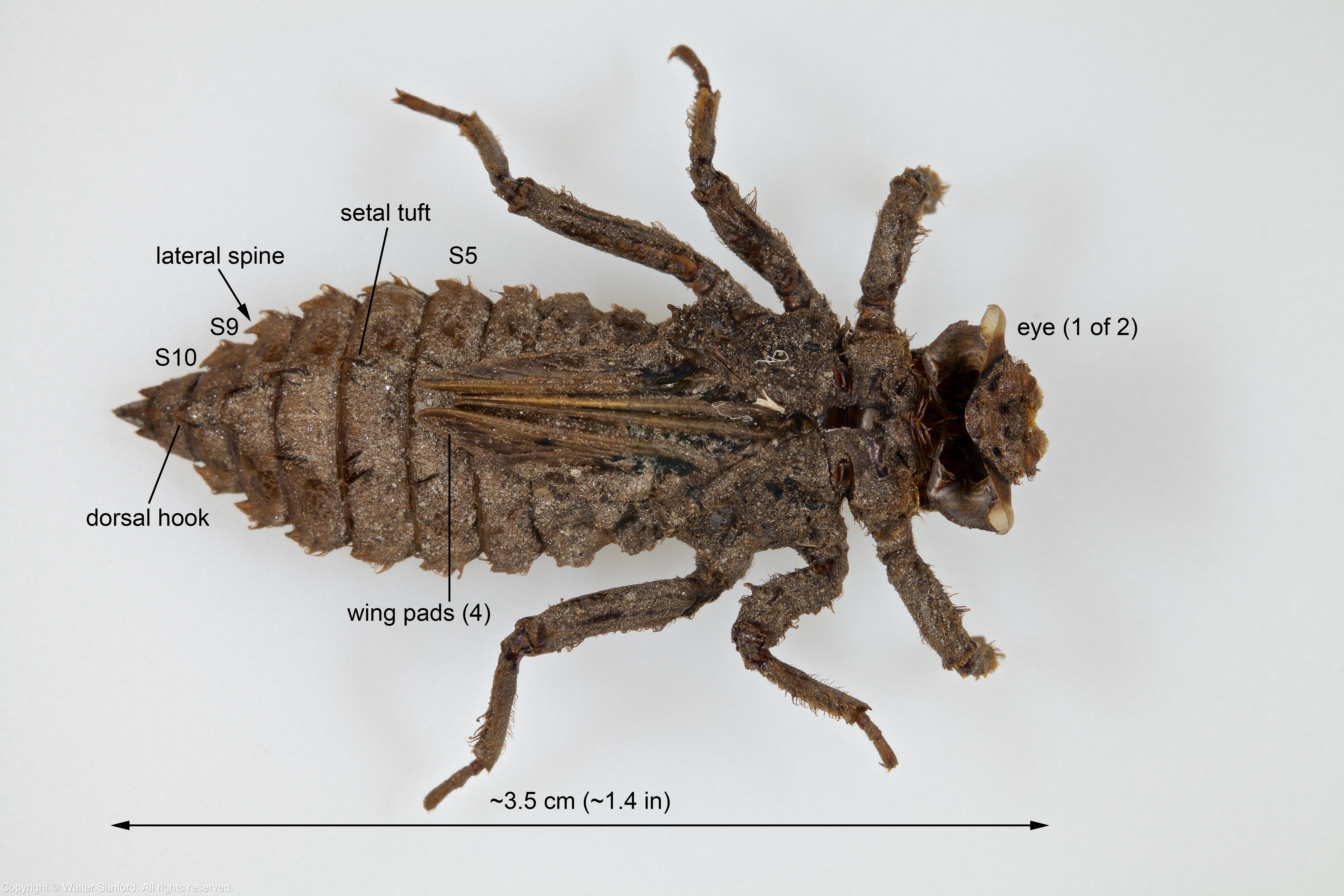
(See a full-size version of the original photo, without annotation.)

A leap of faith is required to see the small dorsal hooks present on abdominal segments two through nine (S2 – S9), but they are there.

Notice the divergent wing pads. The white filaments that extend from the split in the thorax (as shown above) are breathing tubes, artifacts of the unique respiratory system of dragonfly nymphs.

2(1). Lateral spines present on abdominal segments 6-9 only; femora with long, hair-like setae [designatus] [Eastern Ringtail]
2’. Lateral spines present on abdominal segments 5-9 only; femora without long, hair-like setae [constrictor] [Knob-tipped Ringtail]

(See a full-size version of the original photo, without annotation.)

The caudal appendages (terminal appendages) are all about the same length, a key field mark for designatus.

Notice the flat labium that doesn’t cover the face, as shown in the following photo.

This specimen is tentatively identified as an exuvia from an Eastern Ringtail dragonfly (Erpetogomphus designatus). The exuvia that Mike Powell collected is similar in appearance to the following excellent photograph of an Erpetogomphus designatus nymph by Steve Krotzer, Haysop Hill Photography.

Image used with permission from Steve Krotzer.

Related Resource: Odonate Exuviae – a hyperlinked list of identification guides to many species of odonate exuviae from seven families of dragonflies and three families of damselflies.

Tech Tips

The following equipment was used to shoot Photo No. 2 and 4: Canon EOS 5D Mark II digital camera, in manual mode; Kenko 20mm macro automatic extension tubeCanon EF100mm f/2.8L Macro lens (set for manual focus); and Canon MT-26EX-RT Macro Twin LitePhoto No. 1 and 3: Canon EOS 5D Mark II digital camera, in manual mode; Canon MP-E 65mm Macro lens (manual focus only, set for 2x magnification); and Canon MT-26EX-RT Macro Twin Lite. A Sunpak LED-160 Video Light (with a white translucent plastic filter) was used for some photos.

Adobe Photoshop CC 2017 was used to annotate selected images.

Copyright © 2018 Walter Sanford. All rights reserved.

Getting started

February 26, 2018

Michael Powell, a good friend and fellow Northern Virginian, collected several odonate exuviae during a photowalk along the Potomac River in Fairfax County, Virginia USA.

The four exuviae that Mike collected are stored in one of those empty containers of Philadelphia cream cheese spread that I endorsed in a recent blog post. I shot several quick-and-dirty photographs of the exuviae, without removing them from the small plastic tub. Usually I wouldn’t publish photos of this quality. The photos are like a sketch pad that will be used to make a tentative plan for photographing the specimens and attempting to identify them to the genus and species level, if possible.

Spoiler Alert: If you can identify any of the exuviae featured in this post, then PLEASE DON’T TELL ME! I enjoy the challenge of solving the mystery of their identity. Thank you!

Dragonfly exuviae

Mike collected two dragonfly exuviae that are from the Family Gomphidae (Clubtails), as indicated by a flat labium that doesn’t cover the face as well as club-like antennae.

My working theory is the first specimen might be an exuvia from an Eastern Ringtail dragonfly (Erpetogomphus designatus).

Potomac River, Fairfax County, VA | odonate exuvia

Post Update: The identity of the first specimen is confirmed as an Erpetogomphus designatus exuvia.

The next exuvia appears to be a member of the genus Stylurus, possibly plagiatus (Russet-tipped Clubtail).

Potomac River, Fairfax County, VA | odonate exuvia

The preceding photo was focused on the head; the following photo was focused on abdominal segment nine (S9). This specimen may need to be cleaned in order to see more clearly some key field marks used for identification.

Potomac River, Fairfax County, VA | odonate exuvia

Post Update: The identity of the first specimen is confirmed as a Stylurus plagiatus exuvia.

Damselfly exuviae

It is relatively easy to identify damselflies (Suborder Zygoptera) to the family level based upon the shape of the prementum. Both damselfly exuviae that Mike collected are members of the Family Coenagrionidae (Narrow-winged Damselflies).

It is more challenging to identify damselfly specimens to the genus/species level. In this case, possible genera include Argia (Dancers), Enallagma (American Bluets), and Ischnura (Forktails).

Potomac River, Fairfax County, VA | odonate exuvia

The last damselfly exuvia is smaller than the first. It will need to be soaked in soapy water for at least 24 hours in order to make the specimen pliable so it can be re-posed before it is photographed.

Potomac River, Fairfax County, VA | odonate exuvia

Copyright © 2018 Walter Sanford. All rights reserved.

Stenogomphurus rogersi exuvia

February 24, 2018

Bob Perkins collected an unknown species of clubtail larva in February 2017 from a stream located in either Carroll- or Grayson County, Virginia USA. The larva emerged from one of Bob’s holding tanks on 13 March 2017 and metamorphosed into an adult female Sable Clubtail dragonfly (Stenogomphurus rogersi).

A two-step process was used to verify the genus and species of the exuvia.

  1. Determine the family.
  2. Determine the genus and species.

Step 1. Family

First, determine the family of the specimen. For reference, watch the excellent Vimeo video, Identifying dragonfly larva to family (8:06). Here’s the decision tree used to identify the exuvia as a member of the Family Gomphidae (Clubtails).

  • The specimen has a flat labium that doesn’t cover the face (not mask-like). [See Photo No. 2.]
  • Antennae are club-like (not thin and thread-like, as in Aeshnidae). [See Photo No. 1.]
  • Eyes not exceptionally large compared to the size of the head (not large, as in Aeshnidae). [See Photo No. 1.]
(See a full-size version of the original photo, without annotation.)

The white filaments that extend from the split in the thorax (as shown above) are breathing tubes, artifacts of the unique respiratory system of dragonfly nymphs.

Step 2. Genus and species

Gomphidae is the second largest family of dragonflies, so it can be challenging to identify some specimens to the genus and species level.

The dichotomous key for Gomphidae larvae that appears on p. 233 in Dragonflies of North America, Third Edition by Needham et al. was used to verify the genus and species of the exuvia.

dichotomous key: a key for the identification of organisms based on a series of choices between alternative characters. Source Credit: Merriam-Webster Dictionary.

The first couplet [1, 1′] is as follows.

1. Median premental tooth lower than surrounding setae (Fig. 271b); small dorsal hooks present on abdominal segments 8 and 9; lateral spines present on segment 6 (Fig. 272) [rogersi]
1’. Median premental tooth as high as surrounding setae (Fig. 271a); dorsal hooks absent or vestigial on abdominal segments 8 and 9; lateral spines usually absent on segment 6 (Fig. 272) [consanguis]

The following annotated image shows a ventral view of the prementum. Notice the median premental tooth is lower than the surrounding setae.

(See a full-size version of the original photo, without annotation.)

The next annotated image shows a dorsal view of the distal abdomen. A leap of faith is required to see the small dorsal hooks present on abdominal segments eight and nine (S8, S9), but they are there. Also notice the lateral spines present on segment six (S6).

(See a full-size version of the original photo, without annotation.)

Therefore this specimen is confirmed as an exuvia from Stenogomphurus rogersi. Further, the rudimentary ovipositor shown in Photo No. 4 indicates this individual is a female.

(See a full-size version of the original photo, without annotation.)

Bonus Gallery

No. 5 | Sable Clubtail (Stenogomphurus rogersi) | exuvia (head-dorsal)

The Stenogomphurus rogersi exuvia is ~3.0 cm (~1.2 in) long.

No. 6 | Sable Clubtail (Stenogomphurus rogersi) | exuvia (dorsal-lateral)

(See a full-size version of the original photo, without annotation.)

Adult Sable Clubtail dragonflies are slightly larger, on average 4.7 – 5.0 cm (~1.9 – ~2.0 in) long.

Image used with permission from Bob Perkins.

This individual is a female, as indicated by her rounded hind wings and terminal appendages.

Image used with permission from Bob Perkins.

Tech Tips

The following equipment was used to shoot Photo No. 1, 4, 5 and 6: Canon EOS 5D Mark II digital camera, in manual mode; Kenko 20mm macro automatic extension tubeCanon EF100mm f/2.8L Macro lens(set for manual focus); and Canon MT-26EX-RT Macro Twin Lite. A Sunpak LED-160 Video Light (with a white translucent plastic filter) was used for some photos. Photo No. 2 and 3: Canon EOS 5D Mark II digital camera, in manual mode; Canon MP-E 65mm Macro lens (manual focus only, set for 2x magnification); and Canon MT-26EX-RT Macro Twin Lite.

Adobe Photoshop CC 2017 was used to annotate selected images.

Bob Perkins’ photos, taken soon after emergence, were shot using a Canon EOS Rebel T3i camera body and Canon EF-S 60mm macro lens.

Related Resource: Miraculous metamorphosis, a blog post featuring a head-to-head juxtaposition of the same exuvia and dragonfly that are the subjects in this post.

Copyright © 2018 Walter Sanford. All rights reserved.

Miraculous metamorphosis

February 22, 2018

It’s hard to believe an odonate larva that looked like this…

…transformed into the adult dragonfly shown below.

Image used with permission from Bob Perkins.

The Backstory

Bob Perkins, a good friend and fellow Virginian, has been collecting and rearing odonate larvae since 01 January 2017. Bob’s success rate is extraordinarily high, meaning most of the larvae he collects live to emerge as adults.

Bob collected an unknown species of clubtail larva in February 2017 from a stream located in either Carroll- or Grayson County, Virginia USA. The larva emerged from one of Bob’s holding tanks on 13 March 2017 and metamorphosed into an adult female Sable Clubtail dragonfly (Stenogomphurus rogersi).

Amateur- and professional scientists collect and rear odonate larvae in order to observe the adult species that emerge. Then it is possible to reverse-engineer the morphological characters that enable identification of specific species of larvae. An upcoming blog post will feature information about how to identify Sable Clubtail larvae/exuviae.

Tech Tips

The first image is a composite of nine (9) photos taken using the following equipment: Canon EOS 5D Mark II digital camera, in manual mode; Canon MP-E 65mm Macro lens (manual focus only, set for 3x magnification); and Canon MT-26EX-RT Macro Twin Lite.

Adobe Photoshop CC 2017 was used to focus stack the photos and post-process the final output.

The second image was taken by Bob Perkins, soon after emergence, using a Canon EOS Rebel T3i camera body and Canon EF-S 60mm macro lens.

Related Resource: Stenogomphurus rogersi exuvia, a blog post that describes how to identify Sable Clubtail dragonfly larvae/exuviae.

Copyright © 2018 Walter Sanford. All rights reserved.

American Lady butterfly

February 20, 2018

An American Lady butterfly (Vanessa virginiensis) was spotted during a photowalk at Meadowood Recreation Area, Fairfax County, Virginia USA.

This butterfly looks similar to several species with which I’m familiar but isn’t a perfect match, so I consulted the experts on the BugGuide Facebook group. Sincere thanks to Matt Pelikan, Jack Blackford, and Ken Childs for help in identifying the butterfly.

Copyright © 2018 Walter Sanford. All rights reserved.

What is it?

February 18, 2018

It’s time for another exciting edition of “What is it?” Well, what is shown in the following photograph?

What is shown in this photograph?

If you were thinking “empty containers of Philadelphia cream cheese spread,” then you’re only half right.

These small plastic tubs can be repurposed as storage containers for odonate exuviae, such as the Sable Clubtail dragonfly (Stenogomphurus rogersi) exuvia shown below. (Oops, I just noticed it’s time to update the label on the container!)

Storage container for a Sable Clubtail dragonfly (exuvia).

The containers are ideal in many ways. They’re not too big and not too small. The tubs can be “nested” so they don’t take up much space when you’re in the field. For long-term storage, the closed containers can be stacked neatly inside a larger box such as a Rubbermaid Keeper. And the tubs can be used to soak specimens in soapy water in order to clean- and/or re-pose exuviae when they’re pliable.

Finally, think about all the tasty toasted bagels and cream cheese that you get to eat in order to build a collection of specimen containers — that’s what I call a win-win situation!

Copyright © 2018 Walter Sanford. All rights reserved.

Post update: Cordulegastridae exuvia

February 16, 2018

In a recent blog post entitled Cordulegastridae exuvia, I was able to identify the specimen to the family level. Since then, I was able to identify the genus and species.

The dichotomous key for Cordulegastridae larvae that appears on p. 330 in Dragonflies of North America, Third Edition by Needham et al. was used to identify the exuvia.

dichotomous key: a key for the identification of organisms based on a series of choices between alternative characters. Source Credit: Merriam-Webster Dictionary.

The first couplet [1, 1′] is as follows.

1. No lateral spines on abdominal segments 8-9; western [2]
1’. Lateral spines present on segments 8-9; eastern [3]

No. 1 | Cordulegaster sp. | exuvia (ventral)

(See a full-size version of the original photo, without annotation.)

Since the preceding annotated image shows lateral spines on abdominal segments eight and nine (S8, S9), proceed to the third couplet [3, 3′].

3(1’). Palpal setae 4; usually 5 large and 5 small premental setae present; some setae on margin of frontal shelf spatulate (Fig. 391e) [erronea]
3’. Palpal setae 5-7; 5-9 large and 3-5 small premental setae present; all setae on frontal shelf slender, not spatulate (Fig. 391f) [4]

(See a full-size version of the original photo, without annotation.)

The preceding annotated image shows the inner side of the prementum. Four (4) palpal setae are present, plus five (5) large- and five (5) small premental setae. The premental setae on the lower-right side of the prementum seem to be more intact than the ones on the upper-left: the large premental setae are labeled using white numerals; the small premental setae are labeled using red numerals.

The setae on the frontal shelf are mostly missing, as shown below. It’s possible they were broken off either when the larva burrowed in stream sediment (personal correspondence, Sue Gregoire) or when I cleaned the specimen.

No. 3 | Cordulegaster sp. | exuvia (frontal shelf)

(See a full-size version of the original photo, without annotation.)

Genus and species

The number of palpal setae strongly indicates the specimen is an exuvia from a Tiger Spiketail dragonfly (Cordulegaster erronea). Further, the rudimentary ovipositor shown in Photo No. 1 indicates this individual is a female.

The face behind the mask

Do you remember the way the female exuvia looked with its mask-like labium in place? In my opinion, she looked exotically beautiful!

No. 4 | Cordulegaster sp. | exuvia (face-head)

(See a full-size version of the original photo, without annotation.)

Well, that was then and this now. The following photo shows the face and mouth of the exuvia after the face mask was pulled away from the face in order to count the setae on the inner side of the prementum. Look closely at the full-size version of the photo. Yikes, that’s the stuff of nightmares!

No. 5 | Cordulegaster sp. | exuvia (face and mouth)

Related Resources

  • Cordulegastridae exuvia, a blog post by Walter Sanford featuring an exuvia collected by Mike Boatwright.
  • Odonate Exuviae – a hyperlinked list of identification guides to many species of odonate exuviae from seven families of dragonflies and three families of damselflies.

Tech Tips

The following equipment was used to shoot macro Photo No. 2, 3 and 5: Canon EOS 5D Mark II digital camera, in manual mode; Canon MP-E 65mm Macro lens (set for 2x); and Canon MT-26EX-RT Macro Twin Lite. A Sunpak LED-160 Video Light (with a white translucent plastic filter) was used for some photos.

The following equipment was used to shoot macro Photo No. 1 and 4Fujifilm X-T1 digital camera; Fujifilm MCEX-11 extension tube; and Fujinon XF80mm macro lens. An off-camera Fujifilm EF-X500 external flash unit and Sunpak LED-160 Video Light (with a white translucent plastic filter) were used for Photo No. 4. A Raynox DCR-250 close-up filter and Canon MT-26EX-RT Macro Twin Lite was used for Photo No. 1.

Adobe Photoshop CC 2017 was used to annotate selected images.

Copyright © 2018 Walter Sanford. All rights reserved.

Tachopteryx thoreyi exuvia

February 14, 2018

A Gray Petaltail dragonfly (Tachopteryx thoreyi) exuvia was collected on 28 May 2017 by Mike Boatwright in Amherst County, Virginia USA. Gray Petaltail is a member of the Family Petaluridae (Petaltails).

The exuvia has a flat labium, similar to members of the Family Aeshnidae (Darners) and Family Gomphidae (Clubtails). Its seven-segmented antennae are thick and club-like, similar to Clubtail dragonflies.

No. 1 | Gray Petaltail (Tachopteryx thoreyi) | exuvia (face-head)

(See a full-size version of the original photo, without annotation.)

The specimen is ~3.5 cm long and  ~1 cm wide. The wing pads extend to the end of abdominal segment five (S5), as shown in Photo No. 2.

The exuvia features two rows of “setal tufts” (hook-like structures) down its back.

[The] two rows that look like hooks are not hooks at all but rather stout, slight curved setae that are oriented vertically. Source Credit: Ken Tennessen, Western Odonata Facebook group.

(See a full-size version of the original photo, without annotation.)

Also notice a large dorsal hook of abdominal segment nine (S9) overhangs segment 10 (S10).

Photo No. 3 shows a ventral view of the exuvia. Notice the “rudimentary ovipositor” located on abdominal segment nine (S9). An ovipositor is used for egg-laying by all adult damselflies and some species of adult dragonflies: females have this feature; males do not. Therefore, this individual is a female Gray Petaltail.

(See a full-size version of the original photo, without annotation.)

The Backstory

Mike Boatwright has steadfastly resisted my best efforts to lure him to the dark side of odonate exuviae collection and identification. As a concession to me, Mike kindly agreed to look-out for exuviae in unusual habitats. As it turns out, the first exuvia Mike collected for me is a prized specimen. Perhaps I should have titled this post “Mike strikes gold in Virginia!”

Image used with permission from Mike Boatwright.

“Beginner’s luck?” Nope. I know from firsthand experience Mike Boatwright is an extraordinarily keen-eyed odonate hunter. Way to go, Mike!

Related Resources

The dichotomous key for Petaluridae larvae that appears on p. 320 in Dragonflies of North America, Third Edition by Needham et al. is as follows.

1. Antennae 6-segmented, 3rd and 5th segments longer than wide (Fig. 381); cerci each more than 1/2 as long as epiproct; lateral margins of abdominal segments 3-9 not expanded, lateral spines inconspicuous; western [Tanypteryx (p. 322)]
1’. Antennae 7-segmented, 3rd and 5th segments not longer than wide (Fig. 379); cerci each less than 1/2 as long as epiproct; lateral margins of abdominal segments 3-9 expanded, lateral spines conspicuous; eastern [Tachopteryx (p. 321)]

See also Test shots: Tachopteryx thoreyi exuviae, another blog post by Walter Sanford.

Tech Tips

The following equipment was used to shoot all of the preceding photographs: Canon EOS 5D Mark II digital camera, in manual mode; Kenko 20mm macro automatic extension tube; Canon EF100mm f/2.8L Macro lens (set for manual focus); and Canon MT-26EX-RT Macro Twin Lite. A Sunpak LED-160 Video Light (with a white translucent plastic filter) was used for some photos.

Adobe Photoshop CC 2017 was used to annotate selected images.

Copyright © 2018 Walter Sanford. All rights reserved.

How to calculate magnification

February 12, 2018

By definition, a true macro photo is one with a magnification of at least one-to-one (1:1, or 1/1), that is, one unit on the camera sensor is equal to at least one of the same units in the real world. Magnification (in-camera) can be calculated using the following formula.

Mc = size of camera sensor / size of subject on camera sensor

Both measurements must be expressed in the same units in order for the units to cancel during division.

For example, let’s look at the following “full-size” image of a Corduligastridae erronea exuvia. “Full-size” means the image is uncropped (4896 x 3264 pixels).

The photograph shown above was taken using a Fujifilm X-T1 digital camera, Fujifilm MCEX-11 extension tube, and Fujinon XF80mm macro lens. The specifications for the macro lens lists the maximum magnification as 1x. Question is, what is the actual magnification of the subject?

The X-T1 features an APS-C sensor (23.6 mm x 15.6 mm). The dragonfly exuvia is approximately 35 mm in length, or 3451 pixels out of 4896 pixels across the entire image.

Using two equivalent ratios, the following proportion can be used to solve for the length of the exuvia on the camera sensor, in millimeters (mm).

x mm / 23.6 mm = 3451 pixels / 4896 pixels

x = ~16.6 mm. In other words, the exuvia is ~16.6 mm wide on a camera sensor that is 23.6 mm across.

Calculate the in-camera magnification using the following formula.

23.6 mm / 16.6 mm = ~1.4x

The magnification of the macro lens plus extension tube is ~1.4x. 1.19x is listed as the theoretical maximum magnification using an MCEX-11 extension tube mounted on the 80mm macro lens. If we round the spec’d magnification to ~1.2x, the actual magnification of ~1.4x is more than advertised.

Related Resources

Copyright © 2018 Walter Sanford. All rights reserved.

Long-jawed Orb Weavers

February 10, 2018

Long-jawed Orb Weavers (Family Tetragnathidae) are commonly spotted at Mulligan Pond, Jackson Miles Abbott Wetland Refuge, Fairfax County, Virginia USA.

02 AUG 2017 | JMAWR | Long-jawed Orb Weaver

The common name is due to the extended length of the chelicerae (jaws) compared to those of other orb weavers (Araneidae). Source Credit: Family Tetragnathidae – Long-jawed Orb Weavers, BugGuide.

05 SEP 2017 | JMAWR | Long-jawed Orb Weaver

Related Resources

Copyright © 2018 Walter Sanford. All rights reserved.